Story and photo by Thomas Iacobucci
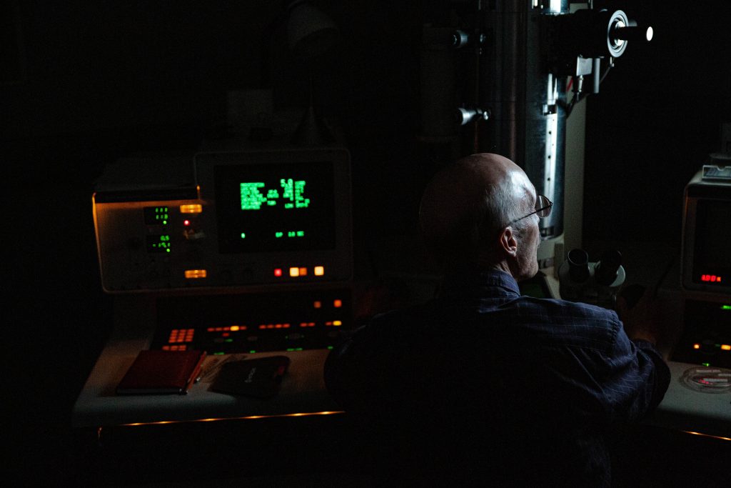
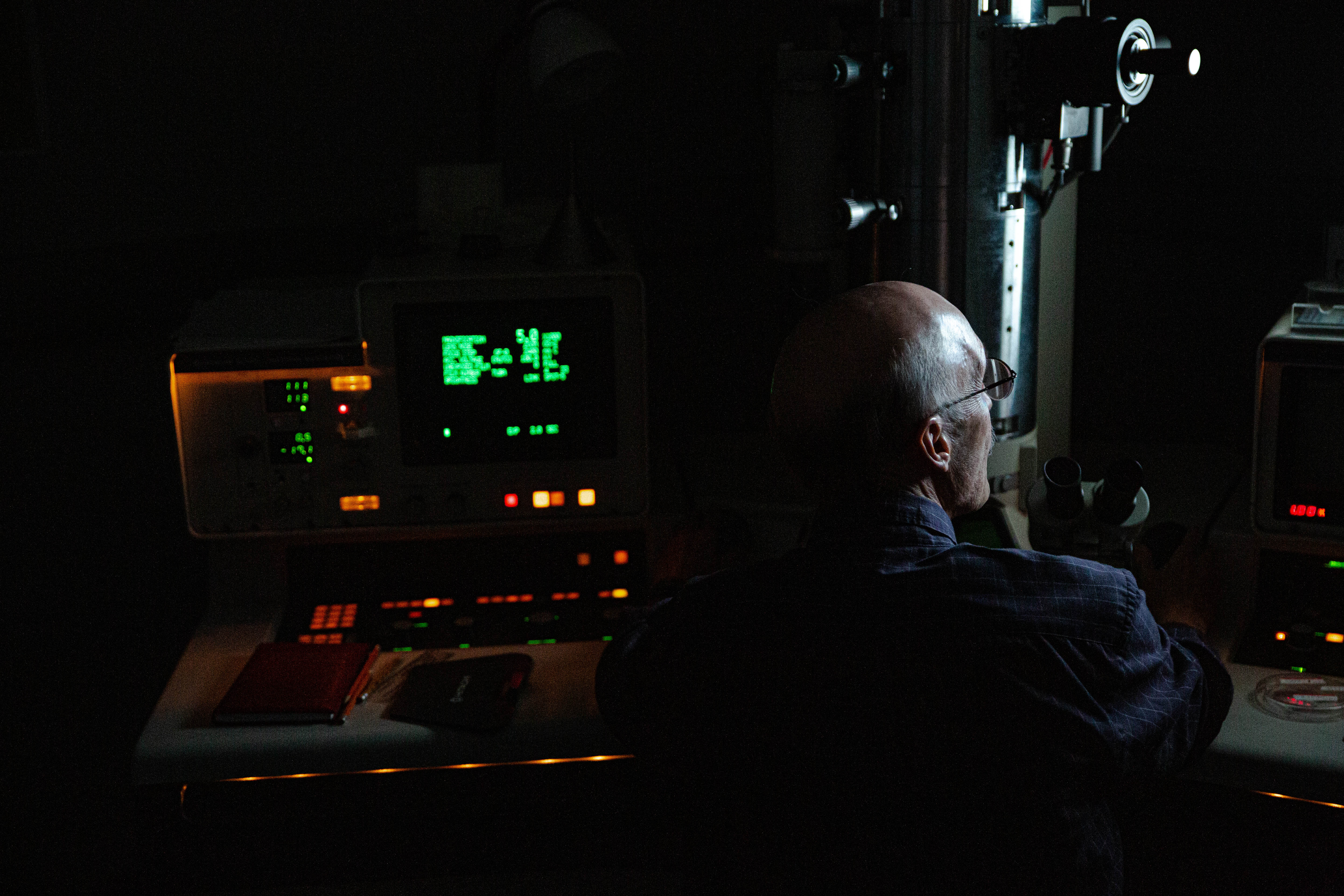
Tony Greco ushered a small group of USF St. Petersburg students into a room no bigger than a closet within the electron microscope facility located on the second floor of the Knight Oceanographic Research Center.
Hitting the lights and ramping up a computer older than most students in the room, Greco took a seat and began to explain what exactly it was the students were looking at.
“We actually have a high-resolution digital camera mounted underneath that we then project up to the monitor,” said Greco, gesturing to a large microscope adjacent to the “IBM-type computer” straight out of the 1980s.
Greco, a professor at the USF College of Marine Science and manager of the electron microscope facility, operated one of five stops during the “Art and Science Night” hosted by the college in conjunction with The Collection, USF St. Petersburg’s art club.
The Nov. 13 event offered attendees the opportunity to walk in the shoes of a marine scientist and see how they view the world.
Of the five stops, participants had the chance to see a virus infecting a cell, look under microscopes at zooplankton and phytoplankton, paint using living bacteria, see the layers of sediment core and manipulate glass to create artistic pieces.
The purpose of the event was for the college to showcase the individual feats that are being studied on a daily basis, and for participating artists to draw inspiration in preparation of an exhibit being held by The Collection on Jan. 8.
“We are going to be doing an exhibition showing some of the work created and inspired by all of this,” said Antonio Permuy, president of The Collection, as he morphed and manipulated a piece of glass during one of the night’s stops.
Though art and science are usually classified as polar opposites, they are one in the same.
“Art is a science of its own, and artists can be inspired by all things nature,” read the Art and Science Night promotional flyer.
Participants were granted a rare look at the artistic side of science, being led from each lab and room by a liaison from the college.
Bespectacled, with eyebrows raised in excitement, Greco began explaining what exactly the small group of huddled students were looking at in his microscopic imaging room.
“The most common thing we’ve looked at are viruses,” said Greco. “Viruses are a little piece of DNA or RNA with a protein coat around them.”
On a more modern monitor, Greco pointed to an image of what he called a T4 virus, or more specifically, an E. Coli cell.
Processing the image on a larger scale allowed students to see the viruses attacking the bacterial cell, telling it to make more viruses and to replicate.
The image on the screen resembled a mid-century abstract-expressionist painting, a perfect rendering of what the artist and students in attendance hoped to see.


